Use and maintenance method of biological microscope
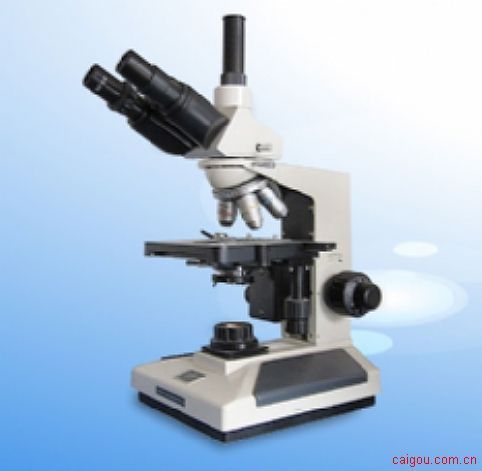
The biological microscope is used to observe and study biological slices, biological cells, bacteria and living tissue culture, liquid sedimentation, etc. At the same time, it can observe other transparent or translucent objects and powders, fine particles and other objects. use: The biological microscope is used for observation of microorganisms, cells, bacteria, tissue culture, suspensions, sediments, etc. by medical and health institutions, universities, and research institutes. It can continuously observe the process of cells and bacteria multiplying and dividing in the culture medium. It is widely used in cytology, parasitology, oncology, immunology, genetic engineering, industrial microbiology, botany and other fields. Binocular biological microscope 44X.3A, Shanghai No.1 Optical Instrument Factory Correct use method and maintenance: 1. When using a microscope, first pay attention to the electrical specifications of the microscope (110V or 220V) and plug in the correct socket. 2. Take out the microscope from the storage box or remove the dust cover. When taking out, you should hold the mirror arm with your right hand and hold the mirror holder with your left hand. After taking out, you should check whether the microscope is dirty. If there is any dirt, you should wipe the lens paper with a little 95% alcohol to remove the dust or contaminants on the objective lens. 3. Turn on and adjust the appropriate light source to prevent the light source from being too high and causing the microbial specimen to dry out. 4. Use a plastic pipette to take out the microorganisms to be observed and place them on a glass slide, and then cover them with a cover glass at a 45-degree angle to avoid air bubbles. 5. Place the finished specimen on the microscope stage and fix the two ends with a slide holder; the operator's left eye is aligned with the eyepiece to observe, adjust the light source and aperture to make the microscope field brightness uniform, usually first The low magnification objective lens starts to be detected as a creature. 6. The glasses are connected to the eyepiece position. Turn the coarse adjustment wheel to make the low-power objective lens almost contact with the specimen slide. After the object appears, turn the fine adjustment wheel to fine-tune it repeatedly until the image is clear. 7. During the microscopic examination, look at the same eyes, look for an appropriate target, and adjust the target to the center of the visual field for observation. 8. Replace the high magnification objective lens to observe, and use the fine adjustment wheel to align the focal length, re-adjust the aperture to the appropriate brightness, and then turn the fine adjustment wheel to repeatedly fine-tune until the object image is clear. 9. During observation, both eyes are opened at the same time to observe with the left eye, and the result with the right eye is depicted. Since the focal length of the high-magnification objective lens is very shallow, it is necessary to turn the fine adjustment wheel on the one hand to describe the result on the one hand. When drawing, there should be no subjective consciousness of the operator, and the observed experimental results should be drawn in full, and cannot be filled in selectively. Binocular biological microscope XSP-8CA O Ring File Folder,Portable O Ring File Folder,Large Capacity O Ring File Folder,Plastic O Ring File Folder TAISHAN JINRI STATIONERY PLASTIC CO.,LTD , https://www.jinri-folder.com